
Diagnosis and treatment
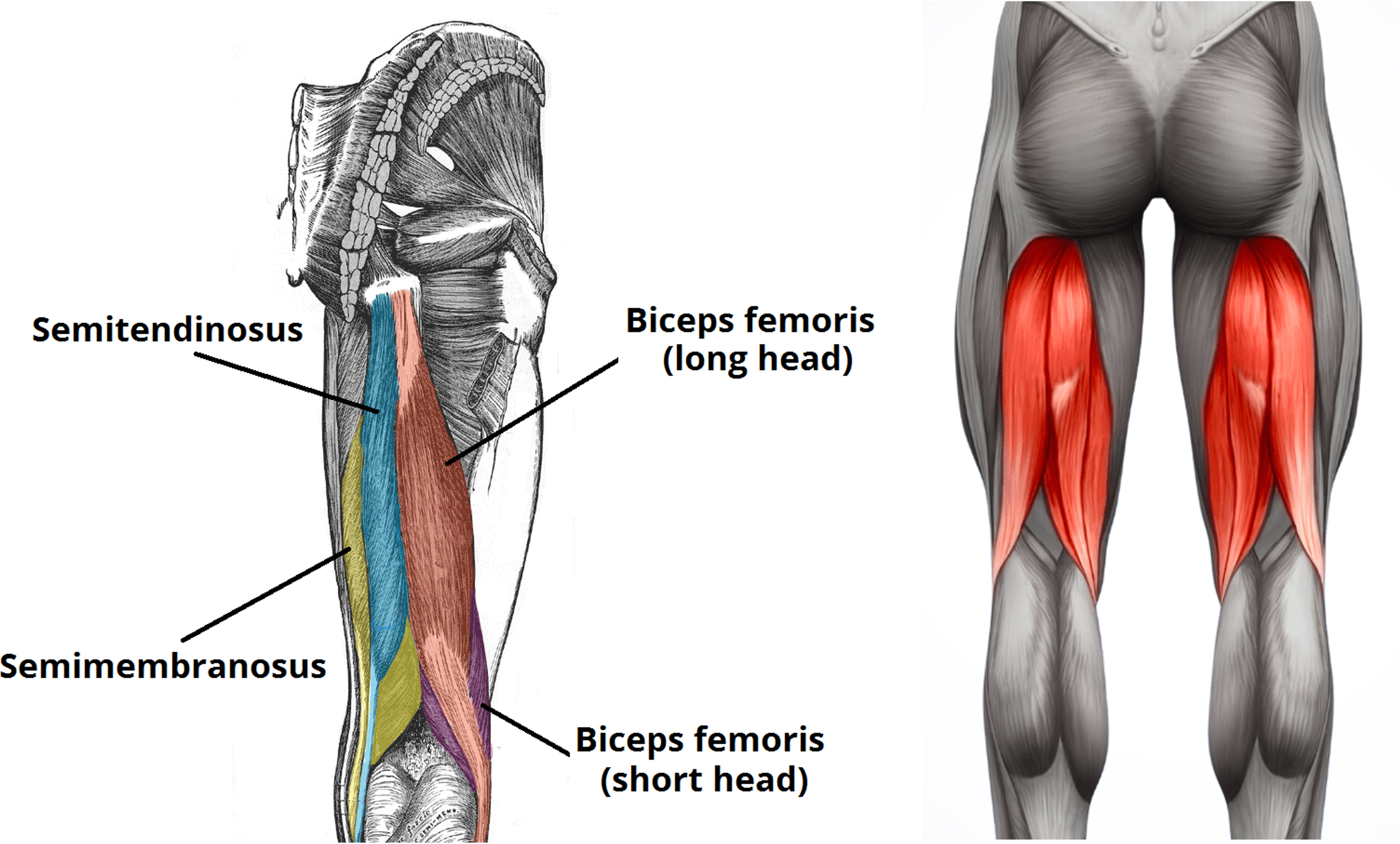
Sports injuries in the proximal thigh (the groin, the front of the thigh, the buttock or the region just below it) are frequently seen in competitive athletes. Injuries of this type involve the muscle itself but most often the tendons of the proximal adductor longus, the hamstrings (biceps femoris, semitendinosus, semimembranosus), or the tendon of the rectus femoris. The sports most affected are football, hockey, athletics and others. The hamstring muscles belong to the posterior compartment of the thigh and comprise the biceps femoris, the semitendinosus and the semimembranosus. They originate at the ischial tuberosity, separate at the midpoint of the thigh and insert at the level of the knee joint. The function of these muscles is essential — extension of the thigh on the trunk, but most importantly flexion of the knee. The biceps femoris also takes part in external rotation, and the other two in internal rotation of the knee. Secondarily, they contribute to stabilising the pelvis and the lower limb.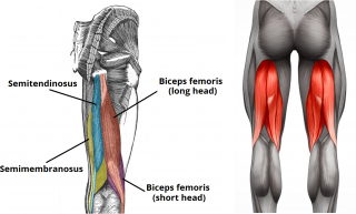 Proximal hamstring injuries can result in a high rate of local morbidity. Injuries can range from strains, partial tears, tendinitis and degeneration through to complete, complex ruptures.
A complete injury means a tear of all three tendons and accounts for approximately 10% of all types of hamstring injury.
Diagnosis:
Clinically, a hamstring injury can produce, at the moment it occurs, intense pain localised at the back of the thigh, below the buttock, and the patient may even hear or feel a pop. In the acute phase a haematoma may be visible in the thigh, and a gap or defect may also be palpable below the buttock. Another clinical sign that may be encountered is the absence of tension in the distal tendons when the patient actively holds the knee flexed at 90 degrees (the bowstring sign).
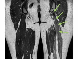
A definitive diagnosis is established with the help of imaging investigations — magnetic resonance imaging (MRI) or musculoskeletal ultrasound.
Proximal hamstring injuries can result in a high rate of local morbidity. Injuries can range from strains, partial tears, tendinitis and degeneration through to complete, complex ruptures.
A complete injury means a tear of all three tendons and accounts for approximately 10% of all types of hamstring injury.
Diagnosis:
Clinically, a hamstring injury can produce, at the moment it occurs, intense pain localised at the back of the thigh, below the buttock, and the patient may even hear or feel a pop. In the acute phase a haematoma may be visible in the thigh, and a gap or defect may also be palpable below the buttock. Another clinical sign that may be encountered is the absence of tension in the distal tendons when the patient actively holds the knee flexed at 90 degrees (the bowstring sign).
A definitive diagnosis is established with the help of imaging investigations — magnetic resonance imaging (MRI) or musculoskeletal ultrasound.
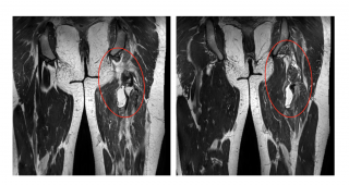 Results at 5 months after surgery:
 Treatment:
The most frequent complaints of patients with a hamstring tear are pain or discomfort and, in competitive athletes, a drop in their level of performance in the sport they play, whether because of persistent muscle weakness or because of pain. Treatment must therefore be tailored to the patient's needs.
Conservative treatment can give good results in patients with a low or moderate level of physical activity, in those with less severe injuries (incomplete, where only a single tendon is affected and retracted less than 2 cm from the ischial tuberosity), or in patients with long-standing injuries. It consists of anti-inflammatory medication, cryotherapy and rest from sport in the first 2 weeks after the injury, followed by a variable period of medical gymnastics at a specialist rehabilitation centre. During this time, corticosteroid injections can be given to patients with persistent symptoms, or injections of regenerative biological products (stem cells from abdominal adipose tissue, PRP or PRF).
Recent studies nonetheless show that the rate of recovery and of patient satisfaction is significantly higher after surgical treatment, at the cost of a potentially longer recovery period, and that the rate of return to sport at the same level as before the injury is as high as 100%. Compared with patients treated surgically, those treated conservatively may also experience sciatic pain if scar tissue at the injury site irritates or encases the sciatic nerve, which can lead to a lengthy recovery period and a greater need for anti-inflammatories.
Surgical treatment is indicated for complete injuries, avulsions and partial injuries with retraction of more than 2 cm of the affected tendon, in physically active patients and competitive athletes, or in any patient in whom conservative treatment has not given optimal results.
The surgical options are not varied, the most frequent technique being suture and reattachment of the proximal tendon at the ischial tuberosity using specific anchors and high-strength sutures. Other options include endoscopic versus open repair, or the use of an autograft where tendons are very retracted and not suitable for direct reattachment.
The patient is positioned prone, with the knee flexed. The operation does not take long, and the most difficult part is the surgical approach, since the sciatic nerve runs 1 centimetre lateral to the hamstring tendons and the posterior femoral cutaneous nerve approximately 0.7 cm lateral.
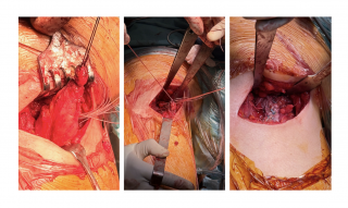
After careful dissection, the tendon identified as torn is cleared of scar tissue, the reattachment site on the ischial tuberosity is debrided, the tendon is sutured with high-strength sutures and reattached using specific anchors or specific interference screws.
Treatment:
The most frequent complaints of patients with a hamstring tear are pain or discomfort and, in competitive athletes, a drop in their level of performance in the sport they play, whether because of persistent muscle weakness or because of pain. Treatment must therefore be tailored to the patient's needs.
Conservative treatment can give good results in patients with a low or moderate level of physical activity, in those with less severe injuries (incomplete, where only a single tendon is affected and retracted less than 2 cm from the ischial tuberosity), or in patients with long-standing injuries. It consists of anti-inflammatory medication, cryotherapy and rest from sport in the first 2 weeks after the injury, followed by a variable period of medical gymnastics at a specialist rehabilitation centre. During this time, corticosteroid injections can be given to patients with persistent symptoms, or injections of regenerative biological products (stem cells from abdominal adipose tissue, PRP or PRF).
Recent studies nonetheless show that the rate of recovery and of patient satisfaction is significantly higher after surgical treatment, at the cost of a potentially longer recovery period, and that the rate of return to sport at the same level as before the injury is as high as 100%. Compared with patients treated surgically, those treated conservatively may also experience sciatic pain if scar tissue at the injury site irritates or encases the sciatic nerve, which can lead to a lengthy recovery period and a greater need for anti-inflammatories.
Surgical treatment is indicated for complete injuries, avulsions and partial injuries with retraction of more than 2 cm of the affected tendon, in physically active patients and competitive athletes, or in any patient in whom conservative treatment has not given optimal results.
The surgical options are not varied, the most frequent technique being suture and reattachment of the proximal tendon at the ischial tuberosity using specific anchors and high-strength sutures. Other options include endoscopic versus open repair, or the use of an autograft where tendons are very retracted and not suitable for direct reattachment.
The patient is positioned prone, with the knee flexed. The operation does not take long, and the most difficult part is the surgical approach, since the sciatic nerve runs 1 centimetre lateral to the hamstring tendons and the posterior femoral cutaneous nerve approximately 0.7 cm lateral.
After careful dissection, the tendon identified as torn is cleared of scar tissue, the reattachment site on the ischial tuberosity is debrided, the tendon is sutured with high-strength sutures and reattached using specific anchors or specific interference screws.
 Postoperative recovery
There is currently no consensus in the literature, but depending on the severity of the injury and the degree of retraction, the patient may be prohibited from bearing weight on the operated limb for up to 6 weeks. A special brace is also used to hold the thigh in extension or the knee in flexion, in order to relax the hamstring muscles. Continuous passive movement can begin 2 weeks after the operation, though this interval can vary with the complexity of the case. The patient can return to sport at least 3 months after surgery.
Proximal ADDUCTOR LONGUS tendon injuries:
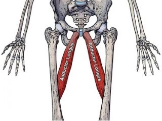
The adductor longus muscle originates at the pubic symphysis and inserts on the inner aspect of the femoral shaft (the linea aspera), its main function being adduction and slight internal rotation of the thigh, as well as stabilisation of the pelvis and the lower limb.
Injury to the adductor longus tendon occurs in athletes through predominantly non-contact mechanisms, during an eccentric load of forced abduction, extension and external rotation of the hip.
Postoperative recovery
There is currently no consensus in the literature, but depending on the severity of the injury and the degree of retraction, the patient may be prohibited from bearing weight on the operated limb for up to 6 weeks. A special brace is also used to hold the thigh in extension or the knee in flexion, in order to relax the hamstring muscles. Continuous passive movement can begin 2 weeks after the operation, though this interval can vary with the complexity of the case. The patient can return to sport at least 3 months after surgery.
Proximal ADDUCTOR LONGUS tendon injuries:
The adductor longus muscle originates at the pubic symphysis and inserts on the inner aspect of the femoral shaft (the linea aspera), its main function being adduction and slight internal rotation of the thigh, as well as stabilisation of the pelvis and the lower limb.
Injury to the adductor longus tendon occurs in athletes through predominantly non-contact mechanisms, during an eccentric load of forced abduction, extension and external rotation of the hip.
 Diagnosis:
A proximal adductor longus tendon tear is characterised by intense pain and an apparent increase in volume, sudden or gradual, on the inner aspect of the proximal third of the thigh or adjacent to the groin. The patient often hears or feels a pop or crack at the moment it happens.
On clinical examination in the acute phase, bruising may be visible either in the groin or lower down the thigh. In the chronic phase, the injury can present as a dull discomfort, made worse by physical activity. The doctor can palpate the torn end of the adductor while it contracts.
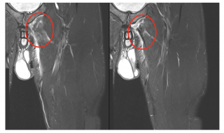
A definitive diagnosis is established following imaging assessment by magnetic resonance imaging (MRI) or musculoskeletal ultrasound.
Diagnosis:
A proximal adductor longus tendon tear is characterised by intense pain and an apparent increase in volume, sudden or gradual, on the inner aspect of the proximal third of the thigh or adjacent to the groin. The patient often hears or feels a pop or crack at the moment it happens.
On clinical examination in the acute phase, bruising may be visible either in the groin or lower down the thigh. In the chronic phase, the injury can present as a dull discomfort, made worse by physical activity. The doctor can palpate the torn end of the adductor while it contracts.
A definitive diagnosis is established following imaging assessment by magnetic resonance imaging (MRI) or musculoskeletal ultrasound.
 Treatment:
The patient's most frequent complaint is a drop in performance and pain during the sport they play. Treatment must therefore be personalised, with recommendations made according to the patient's needs.
Conservative treatment is recommended for all patients with minimal and moderate physical demands. It consists initially of physical rest, appropriate anti-inflammatory treatment, cryotherapy and compression of the thigh using a strap or elastic bandage, followed of course by medical gymnastics at a specialist rehabilitation centre.
For patients who opt for conservative treatment, the literature shows that injections of growth factors with regenerative power, such as stem cells from abdominal adipose tissue, PRP, PRF and others, can be beneficial in repairing incomplete injuries.
Conservative treatment can give good results even in competitive athletes, but no one can guarantee complete recovery.
In any case, it frequently happens that patients continue to complain of residual discomfort and a drop in performance. At that point the possibility of orthopaedic surgery comes into consideration.
Surgical treatment:
Recent studies maintain that surgical treatment leads to a return to sport at the same capacity as before the injury, at the cost of a longer postoperative recovery compared with conservative treatment.
The main indications for surgical treatment include proximal avulsions, high-grade tears, and chronic injuries with residual muscle weakness or compromised function.
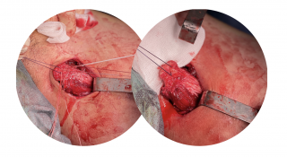
The surgical approach is extremely important and is the hardest part of the operation. It requires a very good knowledge of the anatomy of the groin, since essential vascular and nerve structures pass through this area (the femoral vein, artery and nerve). Through a minimally invasive incision centred on the pubic symphysis and extended along the adductor longus muscle, the torn end of the tendon is identified, scar tissue is debrided and the tendon is prepared for reattachment.
Reattachment can be carried out with special anchors and high-strength non-absorbable sutures, or with interference screws.
The whole operation takes a maximum of 1 hour. Although it is a relatively short operation, it requires a well-prepared surgical team, very good anatomical knowledge and reliable implants with which the surgeon is familiar.
Treatment:
The patient's most frequent complaint is a drop in performance and pain during the sport they play. Treatment must therefore be personalised, with recommendations made according to the patient's needs.
Conservative treatment is recommended for all patients with minimal and moderate physical demands. It consists initially of physical rest, appropriate anti-inflammatory treatment, cryotherapy and compression of the thigh using a strap or elastic bandage, followed of course by medical gymnastics at a specialist rehabilitation centre.
For patients who opt for conservative treatment, the literature shows that injections of growth factors with regenerative power, such as stem cells from abdominal adipose tissue, PRP, PRF and others, can be beneficial in repairing incomplete injuries.
Conservative treatment can give good results even in competitive athletes, but no one can guarantee complete recovery.
In any case, it frequently happens that patients continue to complain of residual discomfort and a drop in performance. At that point the possibility of orthopaedic surgery comes into consideration.
Surgical treatment:
Recent studies maintain that surgical treatment leads to a return to sport at the same capacity as before the injury, at the cost of a longer postoperative recovery compared with conservative treatment.
The main indications for surgical treatment include proximal avulsions, high-grade tears, and chronic injuries with residual muscle weakness or compromised function.
The surgical approach is extremely important and is the hardest part of the operation. It requires a very good knowledge of the anatomy of the groin, since essential vascular and nerve structures pass through this area (the femoral vein, artery and nerve). Through a minimally invasive incision centred on the pubic symphysis and extended along the adductor longus muscle, the torn end of the tendon is identified, scar tissue is debrided and the tendon is prepared for reattachment.
Reattachment can be carried out with special anchors and high-strength non-absorbable sutures, or with interference screws.
The whole operation takes a maximum of 1 hour. Although it is a relatively short operation, it requires a well-prepared surgical team, very good anatomical knowledge and reliable implants with which the surgeon is familiar.
 Postoperative recovery:
Immediately after surgery the patient will be able to bear partial weight on the operated limb, with the aid of two crutches. Anti-inflammatory treatment and cryotherapy are started and medical gymnastics begins immediately. The patient must avoid abduction and extension of the thigh for approximately 1 month, to allow time for the tendon to integrate with the pubic symphysis.
A return to sport is possible at any time after a period of intensive rehabilitation, but no earlier than 12 weeks.
References:
Postoperative recovery:
Immediately after surgery the patient will be able to bear partial weight on the operated limb, with the aid of two crutches. Anti-inflammatory treatment and cryotherapy are started and medical gymnastics begins immediately. The patient must avoid abduction and extension of the thigh for approximately 1 month, to allow time for the tendon to integrate with the pubic symphysis.
A return to sport is possible at any time after a period of intensive rehabilitation, but no earlier than 12 weeks.
References:
- Luengo-Alonso G, Alcobía-Díaz B, Aramberri M, León-Gómez E, Villalón-González L, Valentí-Nin JR. Management of Proximal Hamstring Injuries: Non-operative and Operative Treatment. Curr Rev Musculoskelet Med. 2024 Jul 16;17(9):373–385. doi: 10.1007/s12178-024-09911-0.
- Fletcher AN, Cheah JW, Nho SJ, Mather RC. Proximal Hamstring Injuries. Clin Sports Med. 2021 Apr;40(2):339–361. doi: 10.1016/j.csm.2021.01.003.
- Bertiche P, Mohtadi N, Chan D, Hölmich P. Proximal hamstring tendon avulsion: state of the art. J ISAKOS. 2021 Jul;6(4):237–246. doi: 10.1136/jisakos-2019-000420.
- Azar FM, Canale ST, Beaty JH, editors. Campbell's operative orthopaedics. 14th ed. Philadelphia: Elsevier; 2020.

Nu există comentarii încă. Fii primul!