
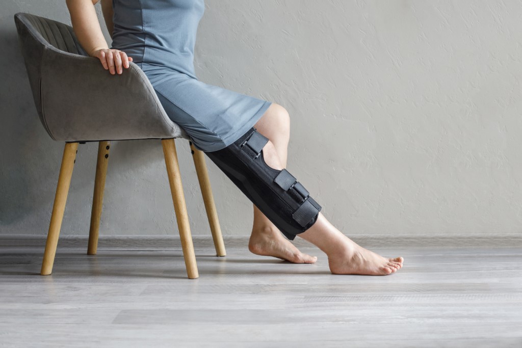
The anatomy of the knee: the role of the meniscus and the anterior cruciate ligament
The knee is a joint made up of the femur, the tibia and the kneecap, plus articular cartilage, the menisci, ligaments and tendons. The meniscus is a fibrocartilaginous structure located between the femur and the tibia. Each knee has two menisci: medial and lateral. These structures play an essential role in absorbing shock and in distributing weight evenly during walking and other activities. The meniscus works as a natural shock absorber, reducing the impact of forces on the articular cartilage and so preventing damage to it. It also helps to stabilise the knee joint, maintaining the balance between the bone surfaces and contributing to the mobility and correct functioning of the joint. The anterior cruciate ligament (ACL), on the other hand, is one of the four main ligaments of the knee. It lies inside the joint and is essential to the stability of the knee during rotation and rapid changes of direction. The ACL prevents excessive movement of the tibia relative to the femur and plays a crucial role in stabilising the joint during high-intensity activities such as running or jumping.The causes and consequences of meniscal and ACL injuries
Injuries to the meniscus and the anterior cruciate ligament can occur for a variety of reasons. The most common causes are sudden, uncontrolled movements such as twisting, jumping and abrupt stops. Sports involving rapid changes of direction, such as football, basketball or skiing, significantly increase the risk of knee injury. Meniscal injuries are caused by a sudden twist of the knee while the foot is planted on the ground. This mechanism can lead to a fissure or even a tear of the meniscus. The patient feels severe pain, swelling and restricted movement of the knee. Without appropriate treatment, the patient may experience long-term pain with limitation of activity. ACL injuries are often the result of a direct impact or of a rotational or hyperextension movement that places too much pressure on the anterior ligament. Once torn, the ACL cannot heal on its own and requires medical intervention. The consequences of such an injury include chronic instability of the knee, difficulty bearing weight on the affected leg, and an increased risk of developing other problems in the knee, including meniscal and cartilage injuries.How to prevent knee injuries
Preventing meniscal and ACL injuries is essential, particularly for those who play contact sports or take part in intense physical activity such as skiing or tennis. Here are some effective preventive measures:- Strengthening the muscles – Strong muscles around the knee, particularly the quadriceps and hamstrings, help to stabilise the joint and reduce the risk of injury. Strength and stretching exercises are essential in preventing injury;
- Warming up properly – Before any intense physical activity it is important to warm up properly, to prepare the muscles and ligaments for effort. Warming up increases blood flow and improves the elasticity of the joints;
- Correct technique – In sports that involve sudden movements and twisting, it is important to use the correct technique to minimise the risk of injury. Training to improve agility and coordination can be a great help;
- Appropriate equipment – Using suitable footwear that provides support and is adapted to the type of surface the sport is played on can considerably reduce the risk of injury.
Diagnostic methods
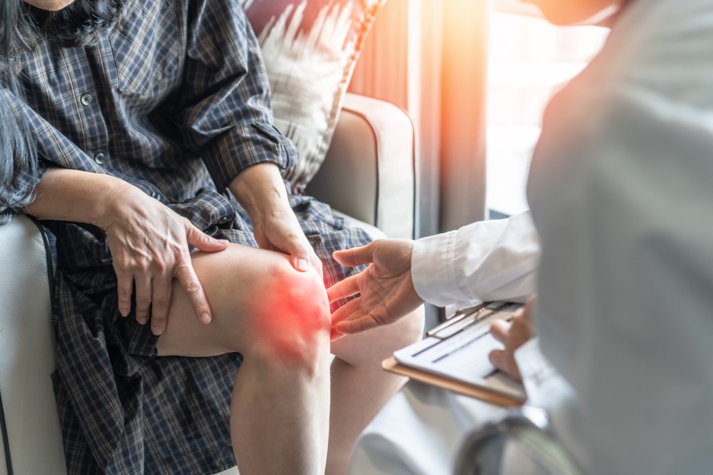 Correctly diagnosing meniscal and ACL injuries is essential in determining the best treatment. The doctor will begin with a thorough clinical examination and will assess the patient's knee: swelling, instability and painful points. The clinical examination includes specific tests, such as the McMurray test for the meniscus and the Lachman or Pivot Shift test for the ACL.
For a more detailed assessment, imaging methods are recommended:
Correctly diagnosing meniscal and ACL injuries is essential in determining the best treatment. The doctor will begin with a thorough clinical examination and will assess the patient's knee: swelling, instability and painful points. The clinical examination includes specific tests, such as the McMurray test for the meniscus and the Lachman or Pivot Shift test for the ACL.
For a more detailed assessment, imaging methods are recommended:
- X-ray – Although a plain X-ray cannot show the meniscus or the ligaments, it can help rule out other problems, such as bone fractures;
- Magnetic resonance imaging (MRI) – MRI is the most effective method for diagnosing meniscal and ACL injuries. It provides clear images of the soft tissues, including the ligaments and cartilage, allowing precise visualisation of the injury;
- Ultrasound – Although not frequently used to diagnose ligament or meniscal injuries, ultrasound can be useful in assessing fluid accumulation in the knee and in detecting superficial tears;
- Arthroscopy – In cases where the diagnosis remains uncertain, the doctor may recommend arthroscopy, which involves introducing a small camera into the joint to view the internal structures directly. This method can be used both for diagnosis and for treatment.
- CT arthrography – for those who cannot have an MRI.
Treating knee injuries
Conservative (non-surgical) treatment
Non-surgical treatment is usually recommended for minor injuries or for people not involved in intense physical activity. It may include:- Rest and immobilisation – Limiting activity and using a brace or crutches can help protect the knee and prevent the injury from worsening;
- Physical therapy – Rehabilitation exercises can help strengthen the muscles around the knee and restore mobility;
- Anti-inflammatory medication – Used to reduce pain and swelling following the injury.
Surgical treatment
In severe cases, particularly when the ACL is completely torn or the meniscus is badly damaged, surgery may be necessary. The most common procedures include:- Meniscal repair
- ACL reconstruction
Rehabilitation and recovery after injury
Recovery after a knee injury, whether to the meniscus or the ACL, requires a well-structured plan. This is an essential part of the healing process, as it helps restore the function of the joint and prevent further complications. Rehabilitation after a meniscal or ACL injury can take from a few weeks to a few months, depending on the severity of the injury and the type of treatment applied (surgical or conservative). After surgery the recovery period is longer and may require a more complex approach. A rehabilitation programme usually includes several stages:- The initial phase (protection and pain control) – Immediately after the injury or surgery, the emphasis is on reducing swelling and protecting the affected joint. In this phase it is important to use braces or crutches and to limit movement of the knee to prevent overloading;
- The mobility recovery phase – As the swelling subsides, the physiotherapist will introduce exercises to restore mobility to the knee. Gentle flexion and extension exercises help prevent stiffness in the joint;
- Strengthening the muscles – In this phase the emphasis is on strengthening the muscles around the knee, particularly the quadriceps and hamstrings. Strong muscles provide additional support to the joint and help prevent recurrence;
- Returning to physical activity – Once muscle strength and mobility have been restored, the patient can begin to gradually resume normal physical activity. It is important to avoid overloading the knee and to follow the recovery programme set by the physiotherapist.

Nu există comentarii încă. Fii primul!