
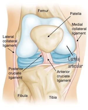
An articular cartilage defect is a common and serious condition of the knee and, as the name suggests, represents damage to the cartilage of the joint. It can appear at any age, either suddenly or over a longer period of time.
Written and medically reviewed by Dr. Ion Bogdan Codorean, senior orthopaedics-traumatology physician. Updated: August 14, 2026.
At least two bones come into contact within any synovial joint. Articular cartilage is the tissue that covers the bony components of the joint.
It has two essential roles:
A cartilage defect is a well-defined area in which the articular cartilage is damaged. Depending on its severity and cause, a cartilage defect can be classified into several grades. The joint most frequently affected by articular cartilage defects is the knee.
Articular cartilage contains no pain receptors, which means the pain is not directly proportional to the extent of the injury. Some cartilage injuries may therefore cause little pain in certain cases. In general, however, the deeper the lesion, the greater the pain.
Symptoms:
Conservative treatment can be applied in the acute phase, or some time after the injury appeared. Acute treatment, for minor and usually post-traumatic injuries, consists of the RICE protocol (Rest, Ice, Compression, Elevation):
If necessary, non-steroidal anti-inflammatory drugs (NSAIDs) can be used to reduce pain and inflammation. Early diagnosis by an orthopaedic surgeon is extremely important.
This is a surgical procedure in which the body is stimulated to produce articular cartilage in the damaged area. In this procedure, the dense surface layer of the bone is penetrated to expose the deeper layers, which contain cells from the red bone marrow.
Advantages: This is the least invasive of all articular cartilage reconstruction techniques, requiring a single operation that can be performed entirely arthroscopically. It is safe, and the results are positive in the majority of patients.
Disadvantages: The new cartilage covering the defect does not have the same structure as normal articular cartilage, and there are concerns about how well it will last over time.
This involves implanting a chondral matrix at the site of the defect, which is then fixed in place with biological products. The chondrocytes (cartilage cells) migrate into this matrix, generating hyaline cartilage with a structure similar to native cartilage.
Advantages: Matrix-assisted chondroplasty is the treatment of choice for large chondral defects. The procedure covers the defect with cartilage of a higher quality than the fibrocartilage generated by microfracture.
As soon as you feel pain, it is advisable to book an appointment with a good orthopaedic surgeon, who will examine you and offer the appropriate solution.
Dr. Codorean offers a precise diagnosis and a personalised treatment plan.