
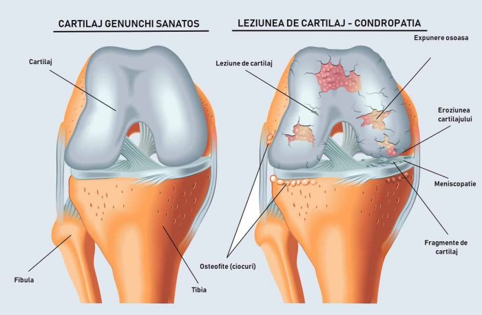
Cartilage covers the bone ends inside a joint. Articular cartilage absorbs shock and ensures minimal friction within the joint. Cartilage injuries are common and their causes are multifactorial. They can appear acutely after trauma, or chronically — as a result of degenerative joint disease, malalignment or joint instability.
Written and medically reviewed by Dr. Ion Bogdan Codorean, senior orthopaedics-traumatology physician. Updated: August 14, 2026.
The most commonly used classification is the Outerbridge system.
The most common symptom of chondropathy (cartilage injury) is pain, though it is not proportional to the grade of the chondropathy. Other symptoms include joint effusion, swelling, clicking and joint locking.
Conservative treatment of chondropathy is used when symptoms are moderate and is based on the RICE protocol (Rest, Ice, Compression, Elevation): rest, ice, compression and elevation of the affected limb above heart level.
Non-steroidal anti-inflammatory drugs may be prescribed for chondropathy. In certain cases, physiotherapy and weight loss may also be recommended.
Several treatment options exist, depending on the surface area of the chondropathy (the cartilage lesion). For grade I–II lesions, treatment is arthroscopic surgery in which the chondral lesions are stabilised, mechanically and thermally, so that synovial fluid can no longer penetrate the fissures in the cartilage.
For grade II–IV cartilage lesions, chondral reconstruction is required. If the affected area is smaller than 1 cm², the microfracture technique is used. This method involves making holes in the bone left without cartilage, down to the layer containing bone marrow cells. The blood emerging from these holes forms a fibrin network that fills the chondral defect.
If the cartilage defect is larger than 1–1.5 cm², other reconstruction techniques are recommended: mosaicplasty, chondral matrix implantation, autologous chondrocyte implantation (ACI) and MACI.
Mosaicplasty involves harvesting autologous cartilage from a minimally load-bearing area of the knee and implanting it into the damaged area.
The preferred technique for chondral reconstruction in grade III–IV chondropathy covering more than 1.5 cm² is the implantation of a chondral matrix (scaffold). Chondrocytes migrate into this matrix and go on to form hyaline cartilage of a higher quality than the pseudo-cartilage generated by microfracture.
Dr. Codorean offers a precise diagnosis and a personalised treatment plan.