
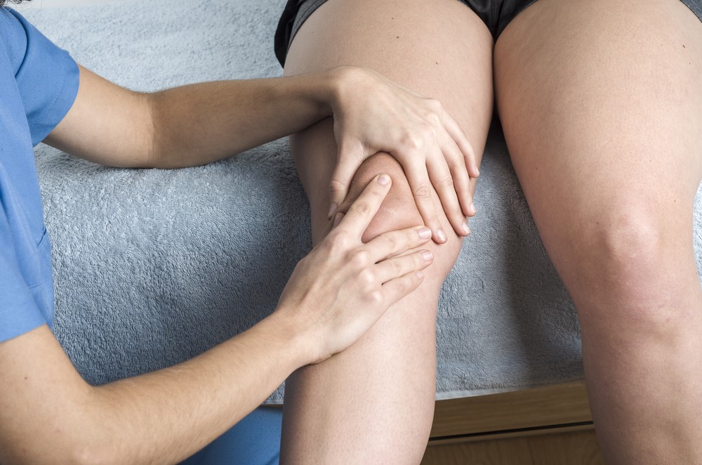
The patella, or kneecap, is a short, flattened sesamoid bone which, together with the tibia and the femur, forms the knee joint. The patella covers the knee, gives it strength and allows it to bend safely. Following a heavy blow or other trauma it can slip to the outside of the knee and cause considerable pain, making it one of the most common knee conditions.
Written and medically reviewed by Dr. Ion Bogdan Codorean, senior orthopaedics-traumatology physician. Updated: August 15, 2026.
Patellar dislocation occurs when the kneecap is pulled out of the femoral groove (the trochlea) as a result of trauma or a heavy blow, but also because of a strong muscle contraction or a fall onto the knee.
More specifically, patellar dislocation occurs in two situations:
The symptoms of a patellar dislocation are:
Patellar subluxation, by contrast, occurs when the kneecap slips slightly and is no longer centred in the femoral groove, but is not completely dislocated, because the joint surfaces remain in contact.
Patellar instability causes the kneecap to track incorrectly in its groove, which damages the cartilage and later leads to osteoarthritis.
The main causes of patellar dislocation are:
In most cases, when the kneecap dislocates it returns to position on its own or is put back by the patient. When this does not happen, the patient must be taken to hospital urgently.
A diagnosis of patellar dislocation is established on the basis of clinical and radiological examination. Investigations include:
Patellar dislocations are usually lateral and can be treated without surgery.
This is recommended for recurrent patellar dislocations or for patellar instability. Depending on the factors responsible for the instability or recurrent dislocation, surgical treatment may consist of:
It is worth noting that the operations listed above can be performed either on their own or in combination.
Whether or not the patient needed surgery to treat the dislocation, rehabilitation is necessary.
After surgery, physiotherapy, kinesiotherapy and massage sessions are recommended to strengthen the joint and muscles, so that the knee regains its mobility and the muscles regain the strength they need. Recovery usually takes several months before the patient is ready for sporting activity.
Rehabilitation involves an individualised set of specific exercises, tailored to the patient's needs, and wearing a brace. To return to sport safely, the patient must pass a number of specific tests.
Dr. Codorean offers a precise diagnosis and a personalised treatment plan.