
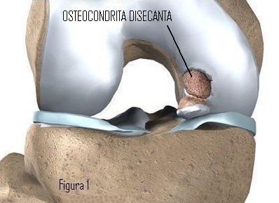
Osteochondritis dissecans is the fragmentation of cartilage and the separation of subchondral bone from a joint surface. It can occur in the knee, but also in other joints such as the ankle. It should not be neglected, as it can be a cause of degenerative joint disease.
Written and medically reviewed by Dr. Ion Bogdan Codorean, senior orthopaedics-traumatology physician. Updated: August 14, 2026.
Osteochondritis dissecans of the knee is a condition in which a fragment of bone covered by cartilage undergoes necrosis. Sometimes it becomes a loose body within the joint, sometimes it collapses, and sometimes only the articular cartilage is damaged. It is also known as a chondral or osteochondral fracture. It usually appears spontaneously, in an active adolescent or a young adult.
Juvenile and adult osteochondritis dissecans involve the same joint defect, but they are separate diseases, because bone metabolism differs considerably with age.
Many doctors believe that juvenile OCD is caused by repeated stress on the bone. Most children with juvenile OCD have played competitive sport from a very young age. A heavy schedule of training and competition can generate stress at the femur that may lead to OCD in children.
The aetiology is uncertain, but it is thought to result from a combination of factors:
The initial symptoms are moderate and worsen over time. Movement in the knee joint becomes painful, and the knee becomes swollen and tender to the touch. Sometimes the pain is severe enough that the patient avoids using the affected leg.
As the disease progresses, the affected portion of bone can collapse, forming a depression in the otherwise smooth joint surface. The cartilage covering this area deteriorates, and the patient feels a catching sensation in the joint, accompanied by pain. In some cases the necrotic bone detaches from the femur and becomes a loose body inside the joint, which can lock the knee.
Conservative treatment may be sufficient in half of all cases of juvenile osteochondritis dissecans. The aim is to promote healing before bone growth stops.
Conservative treatment of the juvenile form can last 10–18 months. During this period it is essential to stop any activity that causes knee pain. The orthopaedic surgeon may recommend using crutches or immobilisation in a plaster splint for several months. As the symptoms subside, rehabilitation exercises can begin under the supervision of a physiotherapist.
It is important to take this condition seriously from the outset and to contact a good orthopaedic surgeon promptly, who can guide you along the right path.
If the affected bone fragment becomes partially or completely detached, surgery is required either to remove the loose body or to fix it in place.
In some cases the orthopaedic surgeon can perform the operation arthroscopically. Using an arthroscope means smaller incisions than open surgery, and recovery and return to activity are also faster.
This is necessary when it is unclear how the fragment can best be positioned, or when the condition is very difficult to treat arthroscopically.
If the loose bone fragment comes from a weight-bearing area, the orthopaedic surgeon will attempt to reattach it wherever possible. Small metal pins or screws are used to fix the fragment. Reattaching the fragment gives considerably better long-term results than removing it.
When the detached fragment cannot be reattached, it is removed. What remains is an articular cartilage defect, which is then treated as such.
Dr. Codorean offers a precise diagnosis and a personalised treatment plan.