
Calcific tendinitis of the shoulder occurs when calcium deposits form on or within the tendons of the shoulder. The tissue around the deposit becomes inflamed, causing significant shoulder pain. The condition is common and most often affects people over 40.
Written and medically reviewed by Dr. Ion Bogdan Codorean, senior orthopaedics-traumatology physician. Updated: August 14, 2026.
There are two different types of calcification in the shoulder: degenerative calcification and reactive calcification.
Wear and tear and the injuries that come with age are the main cause of degenerative calcification. As we grow older, the blood flow reaching the rotator cuff tendons decreases and the tendon becomes weaker. Calcium deposits form in the damaged tendons during the healing process.
Reactive calcification is different. There are three stages of reactive calcification:
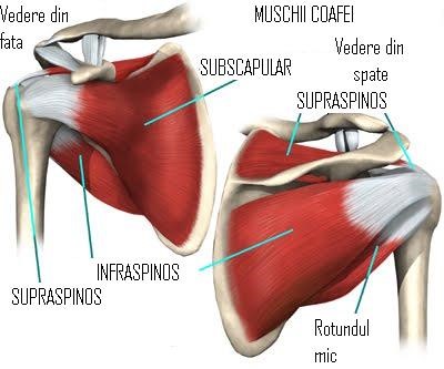
Calcific tendinitis occurs in the rotator cuff tendons. The rotator cuff is a group of tendons connecting the shoulder muscles to the humerus (the upper arm bone). These muscles are called rotators because they rotate the arm at the shoulder joint. Calcium deposits usually appear in the tendon of the supraspinatus muscle.
No one knows exactly what causes calcific tendinitis. Wear and tear, severe injury, ageing, or a combination of these are implicated in degenerative calcification. Some researchers believe calcium deposits form because of insufficient oxygen supply to the tendon. Others believe that pressure on the tendons can cause damage, leading to the formation of calcium deposits. Reactive calcification is an even greater mystery. This type of condition appears in younger patients and seems to resolve spontaneously in many cases.
While the calcium is being deposited, the patient may feel only mild to moderate pain, or may have no symptoms at all. For reasons that are not understood, calcific tendinitis becomes very painful when the deposits are being reabsorbed. The pain and inflammation of calcific tendinitis can limit movement in the shoulder joint. Raising the arm can become painful. At its most severe, the pain can wake the patient from sleep.
The role of a good orthopaedic surgeon is to bring the pain and inflammation under control.
If the pain and restricted movement worsen or affect daily life, the patient requires surgery.
Most operations to treat calcific tendinitis of the shoulder are arthroscopic. The arthroscope is a special camera that can be inserted into the shoulder joint through a small incision in the skin. The orthopaedic surgeon uses the arthroscope to locate the calcium deposits in the rotator cuff. Once found, the deposit is resected (removed) using small instruments and the area is washed out.
In rare cases, open resection is necessary. In open surgery the orthopaedic surgeon removes the calcium deposits by cutting through the surrounding muscles and tissues.
It is very important to see a good orthopaedic surgeon in the early stages of the pain. Delaying brings complications.
Dr. Codorean offers a precise diagnosis and a personalised treatment plan.