
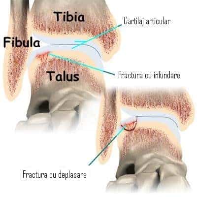
Osteochondritis dissecans of the talus is a condition in which a fragment of bone covered by cartilage undergoes necrosis. Sometimes it becomes a loose body within the joint, sometimes it collapses, and sometimes only the articular cartilage is damaged. It is also known as a chondral or osteochondral fracture.
Written and medically reviewed by Dr. Ion Bogdan Codorean, senior orthopaedics-traumatology physician. Updated: August 14, 2026.
In most cases, osteochondritis dissecans of the talus involves displaced osteochondral fractures. These fractures occur in severe ankle dislocations. The location of the osteochondral fracture is determined by the mechanism of the initial dislocation, and its size and severity vary.
If the bone beneath the cartilage is crushed but the cartilage remains intact, the fragment usually does not displace. If the articular cartilage is torn as well, the fragment can displace. Once a fragment displaces, it loses its blood supply from the bone of origin and becomes necrotic, making spontaneous healing less likely.
There is evidence that a dislocation can destroy the blood supply to the bone over a limited area. The affected area becomes necrotic over time, and this explains cases of osteochondritis dissecans of the talus that appear with no recent history of trauma.
Initially the symptoms are the same as for any ankle dislocation. Swelling, pain and functional impairment appear (an inability to use the affected foot). There are no symptoms that specifically suggest a displaced osteochondral fracture. An X-ray is the best way to diagnose a displaced fracture.
Over time the symptoms worsen. Movement in the ankle joint becomes painful, the ankle swells and becomes tender to the touch. Sometimes the pain is severe enough that the patient avoids using the affected foot. The fragment can cause the ankle to lock.
If the problem is discovered immediately after the ankle dislocation, immobilisation in a cast can be used for six weeks, during which healing may occur. The patient must not put weight on the affected foot and must use crutches throughout the period of immobilisation.
If the affected bone fragment becomes partially or completely detached, surgery is required to remove the loose body. After removal, holes are drilled in the site previously occupied by the fragment, in order to stimulate the formation of new blood vessels and fibrous tissue.
In some cases the orthopaedic surgeon can perform the operation arthroscopically. Using an arthroscope means smaller incisions than open surgery, and recovery and return to activity are also shorter.
This is necessary when the fragment is difficult to reach (the ankle is a small joint and the arthroscope is hard to introduce into certain areas). The incision is usually made at the front of the joint.
Dr. Codorean offers a precise diagnosis and a personalised treatment plan.